


Diagnostic Tools & Technology
Eastside Eye Surgeons uses leading-edge technology for the evaluation and continued care of our patients. Our advanced diagnostic tools include:
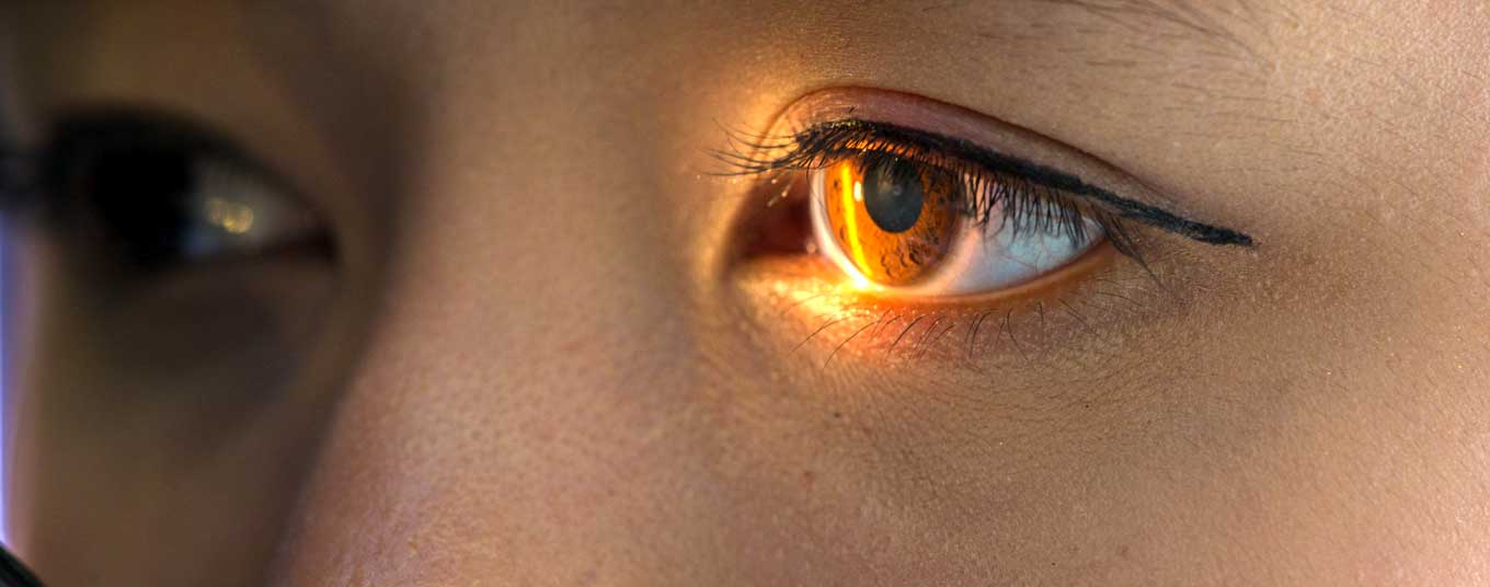
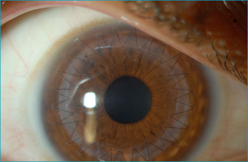
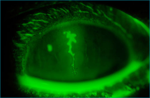
External Photography
Our slit-lamp photography system gives us the ability to document the appearance of your eye with high-quality digital photographs that are available for immediate review. This allows us to compare the progress of corneal and anterior segment conditions in response to treatment. Additionally, these photographs allow us to show you our findings and help further your understanding of your eye conditions.
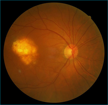
Fundus Photography
Our digital fundus photography system is used to document the status of any retinal or optic nerve conditions. It can be used to follow many diseases such as choroidal nevi (freckles), macular degeneration, diabetic retinopathy and glaucoma.
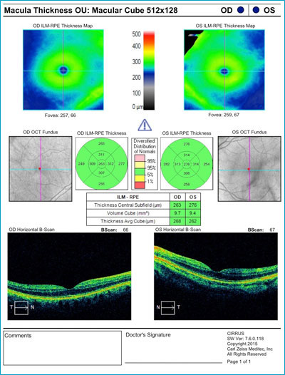
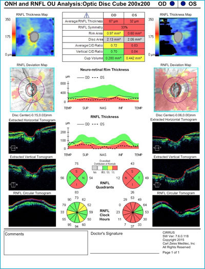
Optical Coherence Tomography (OCT)
The optical coherence tomographer, or OCT, is a cross-sectional imaging modality that uses a laser to scan the retina and optic nerve. The resulting data is processed and analyzed by the computer and displayed as an image that correlates with the microscopic anatomy of the retina and optic nerve. It is a noninvasive test that gives similar information to what might be seen in a microscopic biopsy of the retina or the optic nerve. It can demonstrate fluid under the retina, the status of the nerve fiber layer (damage to which is a hallmark of glaucoma) and the status of the macula (including fluid under the retina and macular degeneration).
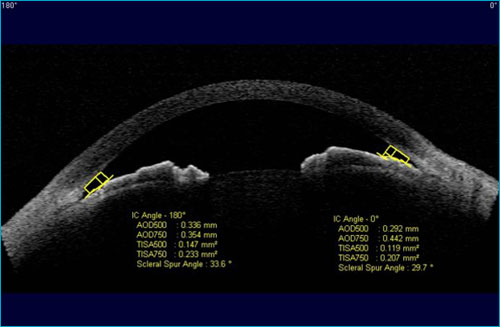
Visante Anterior Segment Optical Coherence Tomography (OCT)
This specialized instrument is used to image the anterior segment of the eye. It provides a detailed scan of the front of the eye to precisely measure the depth of opacities in the cornea while documenting the positioning of LASIK flaps and corneal transplants. Anterior segment OCT is also useful for investigating the anatomy of structures of the eye involved in maintaining pressure, which is important in certain types of glaucoma.
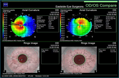
Corneal Topography
Corneal maps, or topographs, are helpful in understanding the shape of the cornea. We diagnose and follow a variety of corneal pathologies, such as keratoconus, through topography. Additionally, corneal topography is a crucial part of our routine exam for all LASIK surgery and premium lens evaluations. Our corneal topographer not only gives us information on the cornea’s surface shape but, when combined with data from the Visante OCT, can be used to provide maps of the cornea’s back surface, as well.
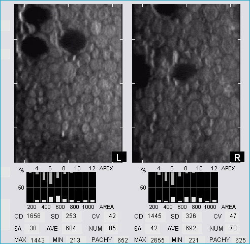
Specular Microscopy
Certain corneal conditions can affect the health of the layer of cells lining the back layer of the cornea. These cells, known as corneal endothelial cells, do not replicate or repair themselves. Conditions such as Fuchs’ corneal dystrophy, prior eye surgery and glaucoma can lead to loss of corneal endothelial cells. If cell count falls below a certain number, corneal swelling occurs, resulting in decreased vision. This is one of the most common reasons for a corneal transplant. The specular microscope images these tiny endothelial cells, which are then computer-analyzed to determine their number and health.
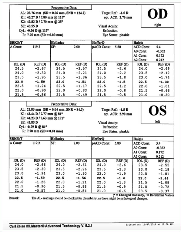
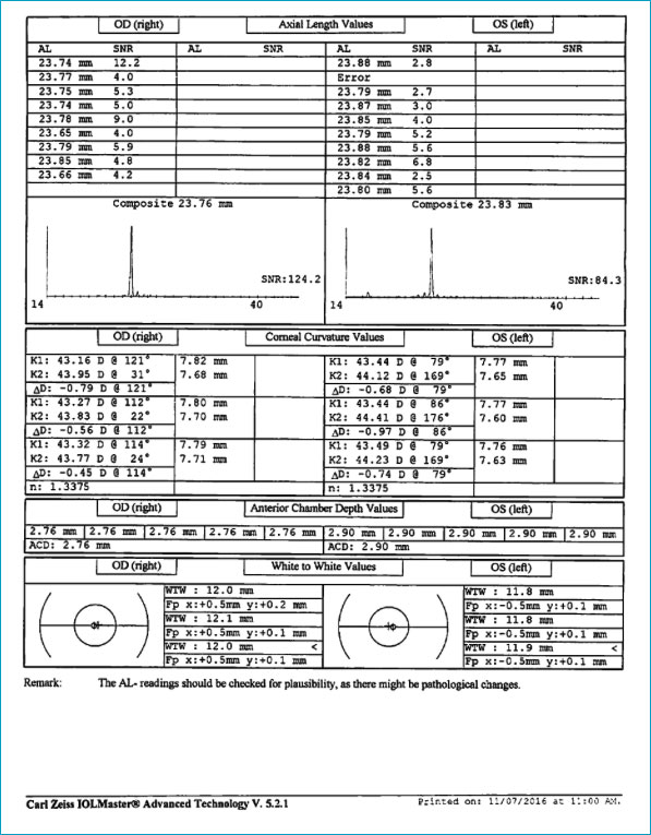
IOL Master & Ultrasound Biometry
Prior to undergoing cataract surgery, your surgeon needs to measure both the shape and length of your eye to determine the optimum power of the intraocular lens to implant. At Eastside Eye Surgeons, we use multiple technologies for each patient in order to get the most accurate and reproducible results. IOL master technology uses a scanning laser to precisely measure the curve and length of your eye. Additionally, manual and ultrasound measurements are taken. Using these multiple modalities helps confirm accuracy and reliability of these critical presurgical measurements.
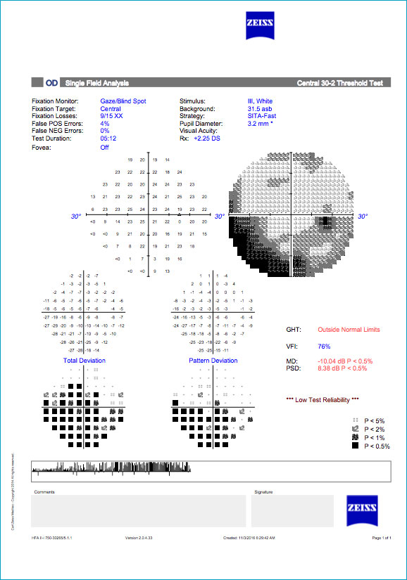
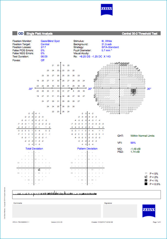
Humphrey Visual Field
Certain medical conditions, such as glaucoma and stroke, or determining potential side effects of certain systemic medications, require monitoring of the visual field. We use a computerized device that tests the central and peripheral portions of your visual field with pinpoints of light. The computer then analyzes the data and provides a variety of statistical information that is useful in monitoring stability or progression and determining effects of treatment.
Learn more about our compassionate, personalized eye care using the most advanced diagnostic tools. Call us at 212-650-0400 for information or use our convenient Request an Appointment form. Patients come to us for diagnosis and treatment from Scarsdale, Chappaqua and Manhattan, New York, Newark, New Jersey, Greenwich, Connecticut and nearby communities.